
What Does the Vitreous Gel Do in the Human Eye?
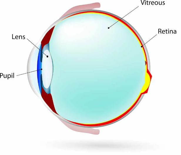
The vitreous gel of the human eye, also called the vitreous humor, is the largest structure within the eye, constituting about 80% of its volume. The vitreous is a clear gel that maintains the shape of the eye and provides a clear space for light pass through to reach the retina. The only structures inside the eye which are not filled with the gel are the lens at the front of the eye, and the retinal lining at the back. Near the center of the retina is the macula, a pigmented region responsible for high-resolution color vision. When light travels through the vitreous humor to the retina and macula, it is then translated to visual information and transmitted by the optic nerve to the brain.
Along with the water which comprises most of the vitreous humor, the gel also contains several types of sugars and salts, collagen fibers, amino acids, and proteins. The gel and fluid allow oxygen and nutrients to flow from the front of the eye to the back of the eye.
As we age, the vitreous humor may begin to shrink due to a decrease in viscosity or thickness. This process is called vitreous degeneration. As the fluid changes from a thick gel-like substance to a thinner liquid consistency, the vitreous humor slowly separates from the retina. This can lead to vitreous floaters, or small disruptions in the visual field such as spots, web-like lines, or rings. No specific treatment is needed in most cases, as the floaters tend to become less noticeable over time.
In some cases, significant vitreous degeneration can lead to detachment of the vitreous from the retina, known as a posterior vitreous detachment (PVD). This can lead to flashes of light and a significant increase in floaters. PVD can also cause blood vessels to stretch and tear, potentially leading to a vitreous hemorrhage. PVD causes traction on the retina, which can lead to several complications such as a retinal tear, retinal detachment, or macular hole.
If any of these conditions arise, treatment is available. This treatment is a surgical procedure called vitrectomy. In a vitrectomy, the vitreous fluid is removed in order to repair the tear, detachment, or hole. After the area has been appropriately treated, the space between the front and back of the eye must be filled. In many cases, it is filled with a gas bubble. Sometimes, silicone oil or a saline-type solution is inserted into the space.
To learn more or to schedule an exam with our doctors, contact Retina Consultants of Nevada at 702-369-0200 or website.